The standing patient position may be better for many patients, but supine or lateral decubitus positions are traditional. There are several different approaches to each kidney and an IMBUS-Advanced physician uses what works best for a patient. Here is a still image of a normal kidney with the areas labeled P for parenchyma, S for renal sinus, and MP for medullary pyramid. The medullary pyramids, when accumulating urine, are seen as small, anechoic areas in the inner parenchyma. The medulla and renal cortex together form a peripheral more hypoechoic area called the parenchyma. With IMBUS, the renal sinus is a single hyperechoic area, except for whatever anechoic urine may be present. The renal columns are portions of the cortex that anchor to the renal pelvis, so the tissue is identical to cortical tissue. The next zone out is the renal medulla, which is made up of the medullary pyramids, separated by renal columns. The central portion, usually called the renal sinus, consists of the calyces, fatty tissue, and the renal pelvis. Here is the posterior anatomy.Ī section through a normal kidney shows three microscopic areas. This posterior long axis view is mostly parasagittal, which means the kidney hilum is always at the medial border. Ultrasound can usually penetrate the posterior tissue and give acceptable views of the kidneys from the back. The kidneys move up and down on these muscles during respiration. Medial to the kidneys are the psoas muscles and directly posterior are the quadratus lumborum muscles, along with adipose tissue.

The liver is cephalad and anterior to the right kidney and the spleen is cephalad to the left kidney as shown in the next image. Ribs cover the upper third of each kidney. Notice in the following image, shown from a lateral view, that the most posterior intercostal space on each side usually gives a long axis, coronal view of the kidneys. The cephalad portion of the kidney is tilted posteriorly. The vessels in this region were covered in a previous chapter but are labeled again in this frontal anatomy diagram. The left kidney may lie more cephalad than the right kidney. However, these beans are not identically shaped and are rotated medially.

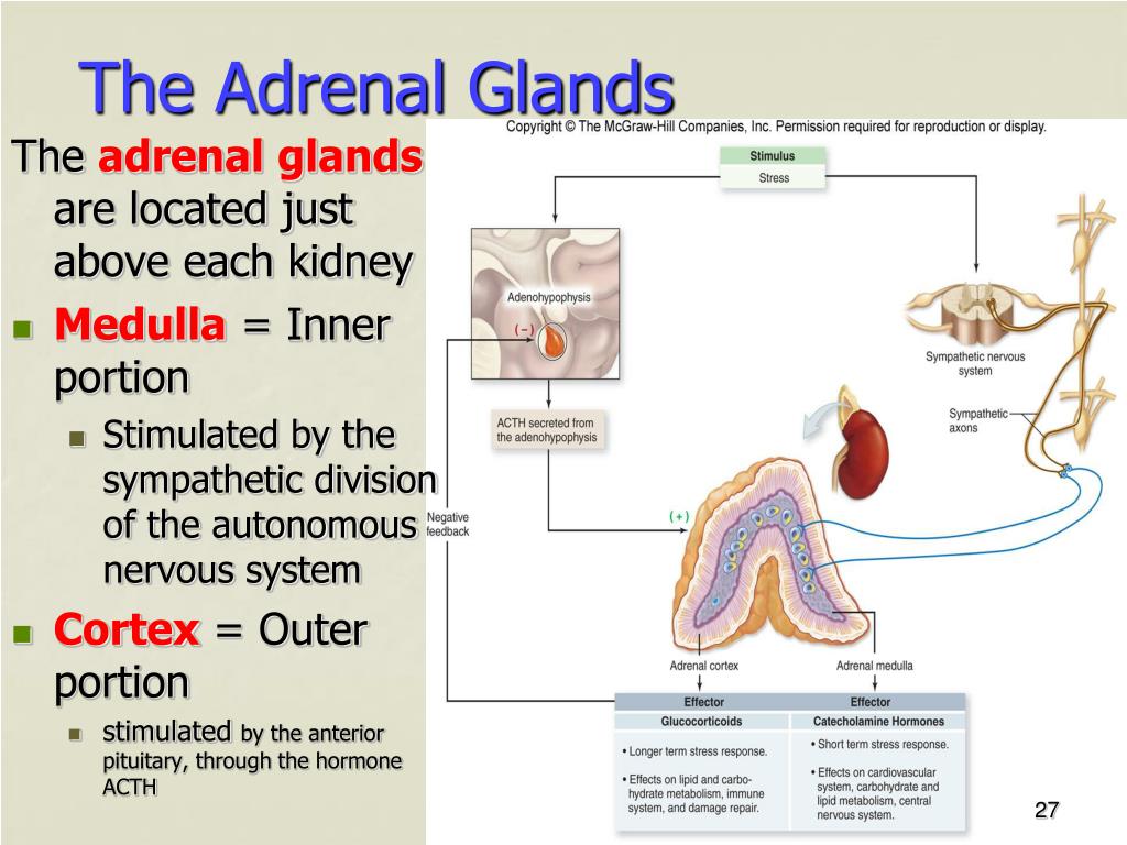
The kidneys lie in the retroperitoneum, behind all other abdominal organs, and really are shaped like “beans”. However, spotting a larger adrenal mass can be an important finding for some patients. Ultrasound is not sensitive for evaluating adrenal glands, so we do not need to worry about increasing the diagnosis of small incidentalomas. The adrenal glands are small and near the kidneys, so large lesions in the adrenal need to be recognized during a kidney exam. However, it is important to identify some additional kidney diseases. IMBUS of the kidneys is most frequently needed in clinic for the question of hydronephrosis.


 0 kommentar(er)
